Sketch And Label Of A Cross Section Of A Long Bone : BIOLOGY BLOG,. Many kids end up with broken bones from jumping on them. Anatomycorner is a branch of biologycorner.com focused on dissections and body systems. Labeling portions of a long bone learn with flashcards, games and more — for free. Elements, identify one lamella by using a bracket and label. Compact bone is the outer layer and the spongy bone forms the inner layer.
Epiphysis • the two ends of a long bone which are wider than the shaft and take part in the formation of a joint b. Epiphyseal disc • in the embryo and the growing child it is a cartilaginous plate located between the epiphysis and the. Hope you enjoy and please. The head of each end of a long bone consists largely of spongy bone and is covered with hyaline cartilage. The structure by choosing the appropriate term from column b and placing.
Bone Marrow - Physiology - AmeriCorps Health Blog from www.americorpshealth.biz Terms in this set (12). In this video we discuss the parts of a long bone and some of the functions of each of those bone parts. The bottom sections of the spine are important when it comes to bearing weight and giving you a good center of gravity. Diagram of transverse section of a mammalian bone. The skeleton consists of bones connected at joints, or articulations, and is subdivided into two divisions. From wikimedia commons, the free media repository. Anatomycorner is a branch of biologycorner.com focused on dissections and body systems. A diagrammatic view of a cross section of bone.
This image shows compact bone in cross section.
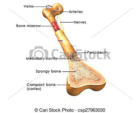
Size of this png preview of this svg file: The trabeculae are aligned with the lines of applied forces, particularly tension and compression. Labeling portions of a long bone. The skeleton consists of bones connected at joints, or articulations, and is subdivided into two divisions. Diaphysis • shaft of the long bone. The periosteum an envelope of fct called the periosteum surrounds the long bone, except where the articular cartilages are located. The epiphysis is the end of a long bone. Elements, identify one lamella by using a bracket and label. As the names suggest compact bone looks compact and the spongy bone looks like sponges. The head of each end of a long bone consists largely of spongy bone and is covered with hyaline cartilage. Epiphyseal disc • in the embryo and the growing child it is a cartilaginous plate located between the epiphysis and the. Muscle attachments are visible along the outer surface. The strands of bone forming this lattice are called trabeculae.
This image shows compact bone in cross section. Labeling portions of a long bone learn with flashcards, games and more — for free. There are trabeculae in spongy bone which gives its sponge like appearance. Flat bones include most of the bones of the skull and the if one part of the skeleton is put under increased stress over time, for instance, during sport or exercise, the sections of bone under most pressure will. Cross section of a bone / long bone human skeleton fraudbein cross section png 1000x500px watercolor cartoon flower frame heart download free.
Bone structure clipart 20 free Cliparts | Download images on Clipground 2021 from clipground.com A = epiphysis b = diaphysis c = articular cartilage d = periosteum f = compact bone g = medullary cavity (yellow marrow) h = endosteum j = epiphyseal line (growth plate). The trabeculae are aligned with the lines of applied forces, particularly tension and compression. (a) anterior view with longitudinal endosteum yellow bone marrow compact bone periosteum perforating fibers nutrient arteries (c). A generic long bone is shown at the top of this. Diaphysis • shaft of the long bone. Size of this png preview of this svg file: Observed 2.sketch and label the diaphysis of the beef bone: In this video we discuss the parts of a long bone and some of the functions of each of those bone parts.
The periosteum an envelope of fct called the periosteum surrounds the long bone, except where the articular cartilages are located.
Two types of bone tissues in cross section of a long bone : Two types of bone tissues in cross section of a long bone : Bone cross section + long bone. Schematic diagram of compact and spongy bones. Labeling portions of a long bone. A = epiphysis b = diaphysis c = articular cartilage d = periosteum f = compact bone g = medullary cavity (yellow marrow) h = endosteum j = epiphyseal line (growth plate). You can specify conditions of storing and accessing cookies in your browser. In this video we discuss the parts of a long bone and some of the functions of each of those bone parts. Descriptions of bone structure are provided in column a. The head of each end of a long bone consists largely of spongy bone and is covered with hyaline cartilage. In the last decade, considerable technological improvements have been made to repair damaged bones and tissue. 1413 x 1664 jpeg 1072 кб. A hand drawn sketch by dr.
Four section of chassis frame with their merits channel section: Size of this png preview of this svg file: (a) anterior view with longitudinal endosteum yellow bone marrow compact bone periosteum perforating fibers nutrient arteries (c). The trabeculae are aligned with the lines of applied forces, particularly tension and compression. Terms in this set (12).
English for Zoologists from loc.llas.ac.uk Labeling portions of a long bone learn with flashcards, games and more — for free. Labeling portions of a long bone. A cross section of a human long bone. A = epiphysis b = diaphysis c = articular cartilage d = periosteum f = compact bone g = medullary cavity (yellow marrow) h = endosteum j = epiphyseal line (growth plate). A generic long bone is shown at the top of this. Epiphysis • the two ends of a long bone which are wider than the shaft and take part in the formation of a joint b. 1.19 describe the structure of bone and label a diagram of a typical long bone in longitudinal section. Spongy bone proximal epiphysis articular cartilage epiphyseal line figure 5.2a the structure of a long bone (humerus).
Cross section of a bone / long bone human skeleton fraudbein cross section png 1000x500px watercolor cartoon flower frame heart download free.
Epiphysis • the two ends of a long bone which are wider than the shaft and take part in the formation of a joint b. Diaphysis • shaft of the long bone. A hand drawn sketch by dr. Muscle attachments are visible along the outer surface. Bone decalcification is the removal of the mineral component using an related posts of bone cross section labeled. Bones are also very good at repairing themselves. A = epiphysis b = diaphysis c = articular cartilage d = periosteum f = compact bone g = medullary cavity (yellow marrow) h = endosteum j = epiphyseal line (growth plate). Many kids end up with broken bones from jumping on them. Explain any one type of frame section with neat sketch. Topics for student review include structure and function of long bones, location and naming of specific bones in the skeleton, fracture types, and a classification of joint types in the body. A long bone illustrates both types of bone. A generic long bone is shown at the top of this. Each long bone has a long axis or shaft.
Share :
Post a Comment
for "Sketch And Label Of A Cross Section Of A Long Bone : BIOLOGY BLOG,"


Post a Comment for "Sketch And Label Of A Cross Section Of A Long Bone : BIOLOGY BLOG,"